The cranial cruciate ligament (CCL) is an important stabilizer in the knee, and ligament tears are common orthopedic injuries seen in pets. If left untreated, significant consequences can occur, but techniques are available to repair damaged CCLs. Our team at Best Friends Veterinary Care provides information about this concerning injury, and options to repair the damage.
Cranial cruciate ligament tears in pets
The knee joint is stabilized by two cruciate ligaments. CCL tears are the most common knee injury in dogs, and occur in cats, but not as frequently. The tear can result from an acute injury to a healthy ligament, but more commonly, the damage is because of slow ligament degeneration. Dogs who have a CCL tear in one knee have a 40 to 60 percent chance of developing the same problem in the other knee, and a partial tear commonly progresses to a full tear over time. Predisposing factors include obesity, poor physical condition, genetics, conformation, and breed. Breeds at higher risk include rottweilers, Labrador retrievers, Akitas, Newfoundlands, and Saint Bernards. Affected pets typically display signs that include:
- Lameness, with varied severity
- Difficulty rising from a sitting position
- Trouble jumping on and off surfaces
- Decreased range of motion in the knee joint
- Decreased activity level
- Swelling along the medial (i.e., the inside aspect) shin bone
Diagnosing cranial cruciate ligament tears in pets
Diagnosis is made by a trained veterinary professional observing your pet’s gait, a physical exam using specific palpation techniques, and X-rays.
- Gait evaluation — Affected pets will limp on the injured limb when walking, and lean away from the limb when standing. When sitting, they will move the injured limb forward, to prevent complete flexion in the knee.
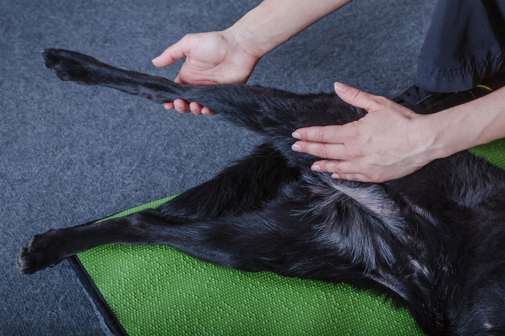
- Physical exam — During the physical exam, swelling around the joint can be appreciated, and clicking noises may be heard when the joint is manipulated. Specific palpation techniques to assess for CCL injury include:
- Cranial drawer test — The veterinarian stabilizes the femur using one hand, and manipulates the tibia with the other. If the tibia moves forward, the CCL is ruptured.
- Tibial compression test — The veterinarian stabilizes the femur using one hand, and flexes the ankle with the other. If the tibia moves forward, the CCL is ruptured. This technique is especially useful in large-breed dogs whose muscular strength and size makes the cranial drawer test difficult.
- X-rays — Imaging the knee by X-ray can confirm joint effusion, evaluate for arthritis, and rule out other issues.
Consequences if cranial cruciate ligament tears in pets are not surgically repaired
A CCL tear causes instability in the knee, which in turn causes movement between the bones and cartilage, resulting in degenerative changes (i.e., arthritis). In some cases, these changes can be observed as soon as one week after the tear occurs. Other consequences of a CCL injury being left untreated include:
- Pain — The initial injury and instability causes the pet significant pain, and as arthritis develops and worsens, the pain will increase.
- Scar tissue — The body tries to stabilize the area by producing scar tissue, which decreases the joint’s range of motion.
- Muscle atrophy — Favoring the injured limb can lead to muscle atrophy.
- Meniscal tear — The continued joint instability can injure the meniscus, causing further pain and instability.
- Weight gain — An inability to exercise can lead to a more sedentary lifestyle, which frequently results in weight gain.
Surgical repair options for cranial cruciate ligament tears in pets
Surgically stabilizing the joint is the best way to help arrest degenerative changes in pets affected by CCL tears. Recommended surgical techniques include:
- Tibial plateau leveling osteotomy (TPLO) — This approach is best for pets who weigh more than 50 pounds, and involves cutting the tibia at the top and rotating the bone so that the femur bears weight on the flat tibial plateau, stabilizing the joint. Typically, exercise is restricted for 8 to 12 weeks, and normal function is usually achieved three to four months after surgery.
- Extracapsular repair — This approach is best for pets weighing less than 50 pounds, and involves passing a large, strong suture behind the knee and through a hole drilled in the tibia, to tighten the joint. Typically, exercise is restricted for 8 to 12 weeks. The suture will break in about 2 to 12 months, but the dog’s own healed tissue should be able to hold the knee at that point.
Enhancing a pet’s recovery after surgery

Strict confinement is crucial in the initial stages after surgery, to help ensure a successful recovery. Your pet will need to be crated, and have only controlled trips to relieve themselves in the first weeks after surgery. Our veterinary professionals will instruct you on gradually returning your pet to exercise over several months. Other factors that can help your pet’s recovery include:
- Joint supplements — Injectable and oral joint supplements can help repair cartilage damage, increase joint lubrication, and decrease inflammation.
- Weight management — Overweight pets are at higher risk for tearing the CCL in the other limb, and more likely to develop arthritis. If your pet is overweight, you should ask our veterinary professionals to devise a weight management plan that is right for your pet.
- Physical therapy — Rehabilitation exercises can help your pet recover more quickly.
CCL tears are painful injuries for pets, but appropriate surgical management can get your pet back on their feet. If your pet is experiencing joint pain, contact our team at Best Friends Veterinary Care, so we can determine the cause of their problem.


Leave A Comment